Low Magnification Micrograph Showing Metastasis Malignant Melanoma Invading Lymph Node — ストック写真

Low magnification micrograph showing a metastasis of a malignant melanoma invading a lymph node. The normal lymphoid tissue has almost completely disappeared and has been replaced by malignant melanoma pigmented and non-pigmented cells. — ストック画像

Low magnification micrograph showing a metastasis of a malignant melanoma invading a lymph node. The normal lymphoid tissue has almost completely disappeared and has been replaced by malignant melanoma pigmented and non-pigmented cells.

 — [著者]の写真 jlcalvo@ucm.es

同じシリーズからのコンテンツ:

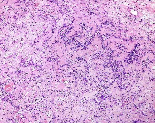
Human spleen amyloidosis. Amyloidosis is caused by abnormal folding of normal soluble proteins leading to fibril formation. These clumps of protein are called amyloid deposits. The spleen is involved in most cases of systemic amyloidosis. The microgr
Human Spleen Amyloidosis Amyloidosis Caused Abnormal Folding Normal Soluble Proteins — ストック写真
Neurilemmomas or schwannoma are rare benign tumours of slow growth derived from the peripheral nerve sheath and composed of perineural Schwann cells. Degenerated or ancient neurilemmomas present typical degenerative characteristics secondary to aging
Neurilemmomas Schwannoma Rare Benign Tumours Slow Growth Derived Peripheral Nerve — ストック写真
Human spleen amyloidosis. Amyloidosis is caused by abnormal folding of normal soluble proteins leading to fibril formation. These clumps of protein are called amyloid deposits. The spleen is involved in most cases of systemic amyloidosis. The microgr
Human Spleen Amyloidosis Amyloidosis Caused Abnormal Folding Normal Soluble Proteins — ストック写真
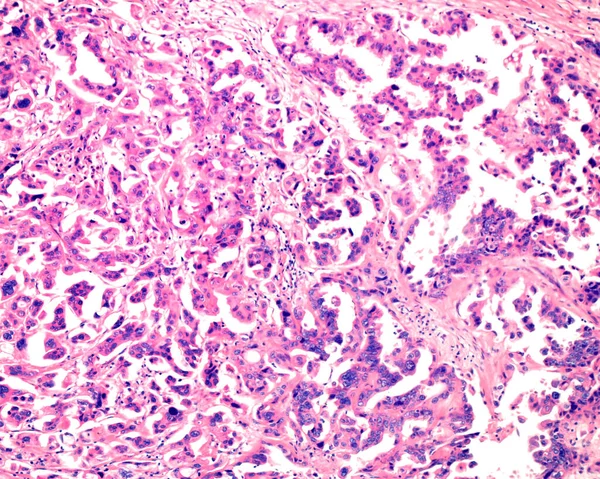
Ovarian mucinous cystadenoma (15-20% of ovarian tumours) is a usually large and benign tumour. But, as shown in this micrograph, it can present cytologic atypia and infiltrative pattern (mucinous cystic adenocarcinoma).
Ovarian Mucinous Cystadenoma Ovarian Tumours Usually Large Benign Tumour Shown — ストック写真
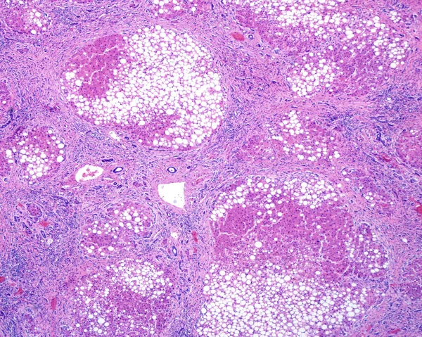
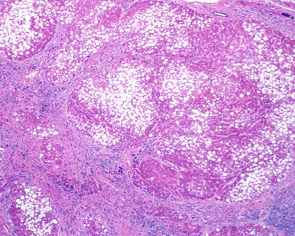
Human liver. Cirrhosis. Low magnification micrograph showing regenerating nodules of hepatocytes (with an extensive fatty change), separated by fibrous septa with chronic inflammatory infiltrates. Cirrhosis of the liver is a result of severe damage t
Human Liver Cirrhosis Low Magnification Micrograph Showing Regenerating Nodules Hepatocytes — ストック写真
Anthracotic リンパ節。石炭粉じん、煙による肺内のリンパ節または汚染にカーボンの蓄積が見つかった最も一般的。H ・ E stai
Anthracotic リンパ節 — ストック写真
Human liver. Drug-induced liver injury by carbon tetrachloride. Light microscope micrograph showing clear areas of degeneration and necrosis of hepatocytes around the central vein with scarce inflammation and some macrophages loaded with pigment.
Human Liver Drug Induced Liver Injury Carbon Tetrachloride Light Microscope — ストック写真
Human spleen amyloidosis. Amyloidosis is caused by abnormal folding of normal soluble proteins leading to fibril formation. These clumps of protein are called amyloid deposits. The spleen is involved in most cases of systemic amyloidosis. The microgr
Human Spleen Amyloidosis Amyloidosis Caused Abnormal Folding Normal Soluble Proteins — ストック写真
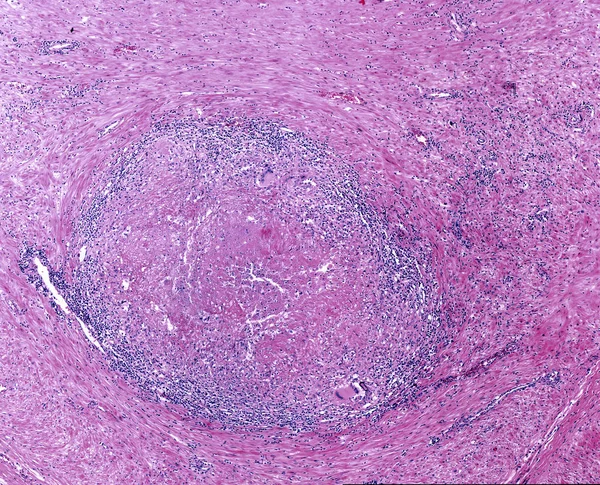
中心壊死 (乾酪壊死)、リンパ球浸潤、多核巨細胞のラングハンス細胞と結核の肉芽腫.
結核の肉芽腫。ラングハンス細胞 — ストック写真
Low magnification micrograph showing a reticulosarcoma, a malignant lymphoma, infiltrating the muscular layer of the stomach.
Low Magnification Micrograph Showing Reticulosarcoma Malignant Lymphoma Infiltrating Muscular Layer — ストック写真
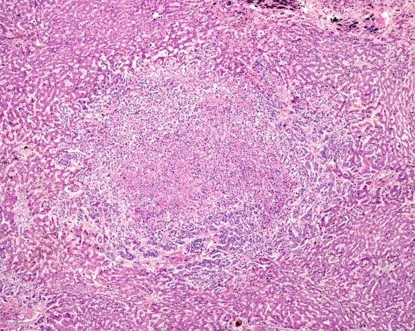
Human liver. Cholangiocarcinoma. Nodule of tumour cells showing central necrosis and inflammatory infiltrates, surrounded by normal hepatocytes.
Human Liver Cholangiocarcinoma Nodule Tumour Cells Showing Central Necrosis Inflammatory — ストック写真
Human liver. Cirrhosis. Low magnification micrograph showing regenerating nodules of hepatocytes (with an extensive fatty change), separated by fibrous septa with chronic inflammatory infiltrates. Cirrhosis of the liver is a result of severe damage t
Human Liver Cirrhosis Low Magnification Micrograph Showing Regenerating Nodules Hepatocytes — ストック写真
人間の肝硬変。肝細胞は、慢性的な炎症細胞の浸潤と線維性結合組織セプタムに囲まれた再生結節で構成されます。茶色の顔料デポも見られています。.
人間の肝硬変 — ストック写真
Human liver. Cirrhosis. Low magnification micrograph showing regenerating nodules of hepatocytes (with an extensive fatty change), separated by fibrous septa with chronic inflammatory infiltrates. Cirrhosis of the liver is a result of severe damage t
Human Liver Cirrhosis Low Magnification Micrograph Showing Regenerating Nodules Hepatocytes — ストック写真
Human spleen amyloidosis. Amyloidosis is caused by abnormal folding of normal soluble proteins leading to fibril formation. These clumps of protein are called amyloid deposits. The spleen is involved in most cases of systemic amyloidosis. The microgr
Human Spleen Amyloidosis Amyloidosis Caused Abnormal Folding Normal Soluble Proteins — ストック写真

利用情報

このロイヤリティフリーの写真「 Low magnification micrograph showing a metastasis of a malignant melanoma invading a lymph node. The normal lymphoid tissue has almost completely disappeared and has been replaced by malignant melanoma pigmented and non-pigmented cells. 」は、標準ライセンスまたは拡張ライセンスに従って、個人的および商業的な目的で使用できます。標準ライセンスは、広告、UIデザイン、製品パッケージなど、ほとんどのユースケースをカバーし、最大500,000部の印刷を許可します。拡張ライセンスでは、無制限の印刷権を持つ標準ライセンスに基づくすべての使用例が許可され、ダウンロードしたストック画像を商品、製品の再販、または無料配布に使用できます。

このストックフォトを購入して、最大3840x3072 の高解像度でダウンロードできます。 アップロード日: 2022年7月3日